Ophthalmic foundation model
One foundation model for every image, every device and every visit.
FUNDUS · OCT · ANGIOGRAPHY · ANTERIOR SEGMENT · TOPOGRAPHY · RECORD
Demand
Americans at risk for retinal disease.
Capacity
Projected US ophthalmology workforce gap by 2035.
Hidden
Of people with an irreversible eye disease don't know they have it.
Time
Of an ophthalmologist's time per patient is spent in the record, not with the patient.
Missed
Of clinically significant cases are missed at routine eye exams.
Patientdataisscatteredacrossdevicesthatdon'ttalktoeachother,andscarcespecialisttimeisspentonrecordsandonpatientswhodidn'tneedtobethere.
Platform
01 · Integrate01 · Unified patient data
Integrate
Every ophthalmic image and the medical record, together in one view.
02 · Augment02 · Decision support
Augment
Physician-in-the-loop decision support from one multimodal model.
The physician always makes the final call.
A device-agnostic intelligence layer that powers multiple clinical products.
Fovea™
Integrated view
Every image and record in one place.
- Vendor-neutral: native data from any device, not screenshots
- One timeline: scans, pressures, medications, diagnoses
- Printouts become trendable data
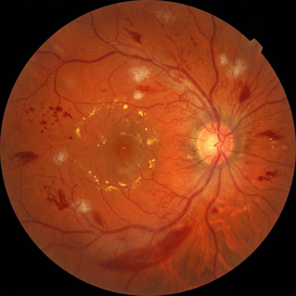
Fundus
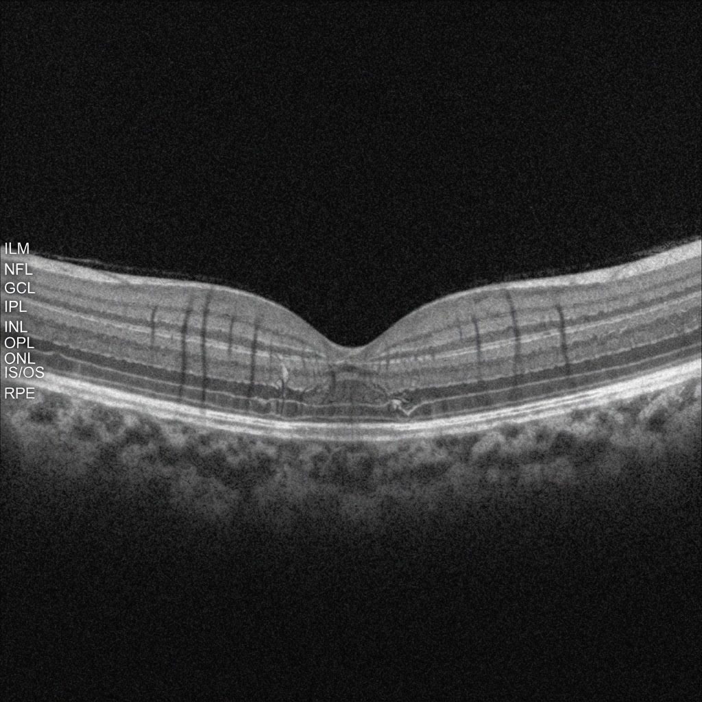
OCT

Visual field
Insight™
Pan-ophthalmic decision support
Multi-disease answers from one image.
- Fundus and volumetric OCT, combined
- Attention maps show where the model looks
- Lesion-level detail, high specificity on healthy eyes
In the clinic
Results
Missed referable disease vs. ophthalmologists
Benchmarked against 10 ophthalmologists from 8 centers, on glaucoma, AMD and diabetic retinopathy. No referable case was caught by the readers and missed by the model.
False positives vs. non-specialist graders
Referable diabetic retinopathy.
One photo, three diseases.
6Diabetic retinopathy
AMD
Glaucoma
Every 100 diabetics screened
Today
100
eye-doctor visits needed, and disease is missed when patients skip them.
With Lumiere
15
eye-doctor visits, and 9+ of 10 diabetic retinopathy cases caught.
Based on published validation. Assumed prevalence: DR 10%, AMD 12%, glaucoma 5%.
From screening to prioritization.
252 patients, a portable handheld camera the model had never seen. No fine-tuning. Image only.
AUC · Referable DR, zero-shot
Unnecessary referrals vs. a clinical-data model
Vision-threatening cases seen within 4 weeks
Vision-threatening DR seen within 4 weeks · demand 25% over capacity
Ranked as Very urgent · Urgent · Priority · Routine. Note: manuscript submitted.
Unnecessary referrals measured at matched 97% sensitivity.
Science
The models behind Lumiere are built on published, peer-reviewed research in glaucoma, retina, OCT and retinal vasculature.
Peer-reviewed papers
Research areas
Artificial intelligence in retinal care: transforming the doctor-patient partnership
How AI is reshaping the relationship between retina specialist and patient, and how to deploy it responsibly.
Shifting the retinal foundation models paradigm from slices to volumes for optical coherence tomography
Reading the full 3D OCT volume instead of a single slice: average AUROC 0.94 vs 0.90 for the best image-based model.
Ophthalmology foundation models for clinically significant age macular degeneration detection
A benchmark of six foundation models on 70,000 expert-labelled fundus images from seven datasets, for moderate-to-late AMD.
GONet: a generalizable deep learning model for glaucoma detection
Trained on more than 119,000 fundus photographs from seven datasets; out-of-distribution AUC 0.88–0.99.
Computerized analysis of the eye vasculature in a mass dataset of digital fundus images: the example of age, sex, and primary open-angle glaucoma
An automated vasculature pipeline applied to 32,768 fundus images; glaucoma shows a pattern consistent with accelerated vascular ageing.
Deep learning generalization for diabetic retinopathy staging from fundus images
DR staging developed on 91,984 images from six independent datasets, designed to generalize across populations.
LUNet: deep learning for the segmentation of arterioles and venules in high resolution fundus images
High-resolution artery/vein segmentation that outperformed three benchmark methods on a local test set and four external test sets.
Leuven-Haifa high-resolution fundus image dataset for retinal blood vessel segmentation and glaucoma diagnosis
An open dataset of 240 expert-corrected artery/vein annotations with glaucoma labels.
FundusQ-Net: a regression quality assessment deep learning algorithm for fundus images quality grading
Automated 1–10 image-quality grading; 99% accuracy on an external test set. It is the quality gate in front of every analysis.
Selected publications · 9 of 16
Live demo
Pick a sample case and run the model. Insight™ reads the image, shows where it is looking and drafts the finding. The physician still makes the call.
Fundus · Left EyeDemonstration on sample data. Not a medical device and not for clinical use.
Whattheeyereveals,weilluminate.
Contact
Clinical collaborations, deployment and integration. Tell us what you’re working on.
contact@lumiere-ai.com